Multimodal Contrast Agents for Brain Diseases and MRI imaging
This project is the extension of the European project LUPAS in which we successfully prepared nanoprobes that could be used for in-vitro, ex-vivo and in-vivo targeting and imaging of amyloids. It focuses on providing new knowledge in chemical design and smart targeting of misfolded protein amyloid lesions in Alzheimer´s disease. Novel molecular insight regarding the pathological hallmarks and the specific chemical requirements for optimal biospecific and biostable amyloid nano-ligands will be achieved.
The theme of research focuses on the design and the study of hybrid (organic-inorganic) nanoparticles as contrast agents for the imaging of brain inflammation associated with pathologies such as ischemic stroke or Alzheimer disease.
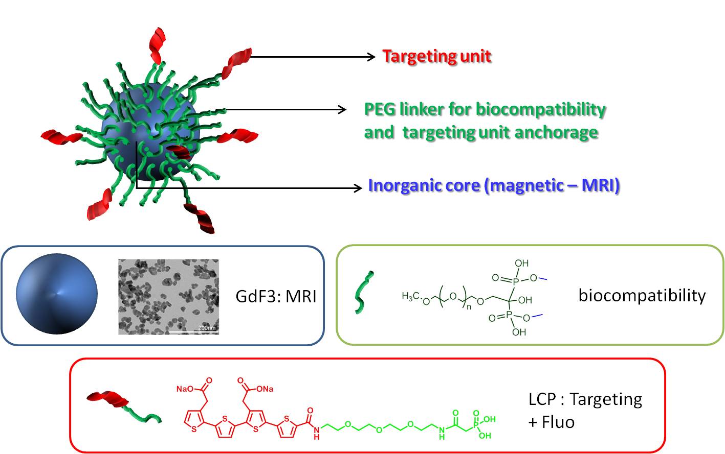
General representation of hybrid nanoparticles used as contrast agents.
These particles consist in a Rare-earth fluoride core (GdF3 for example), exhibiting properties such as magnetism or radio opacity, making them suitable for Magnetic resonance (MR) or CT-scan imaging. Such systems are then functionalized with organic ligands ensuring their biocompatibility and making them suitable for in-vivo injection. Finally surface of the nanoparticles can be modified with specific organic moieties for multiphotons imaging.
Within the frame of a European project (LUPAS), preliminaries studies have shown the potential of GdF3 based hybrid nanoparticles as contrast agents for MRI. After stereotaxic injection of a solution in a mice brain, a negative contrast (T2) is clearly observed
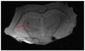
MRI image of a mice brain after nanoparticles injection.
A dynamic follow-up was performed after injection in the vein tail, using florescence imaging. Pictures show the targeting of beta amyloids with a shift of the luminescence from the blood vessel to the brain parenchyma.
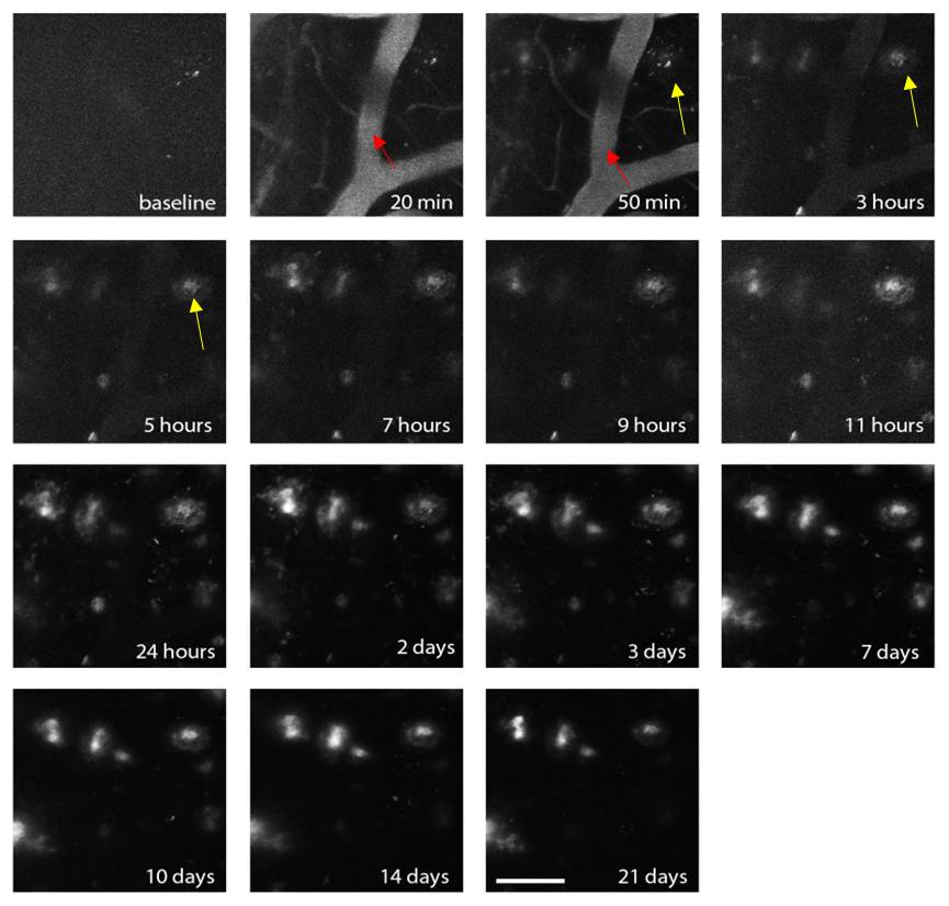
Follow-up of beta amyloids targeting by GdF3 nanoparticles. A shift of the luminescence from the blood vessel to the brain parenchyma is observed.
Several projects concerning that theme are currently ongoing. One of those projects (ANR NANOBRAIN) focuses on the specific targeting and imaging of brain inflammation following reperfusion after stroke. Another project (H2020 SPCCT) is centered on the use of GdF3 nanoparticles as contrast agent for a new imaging modality known as spectral scanner allowing K-edge Imaging, which consists in the accurate detection of gadolinium atoms in the body.
Collaboration
The partners are the University of Linköping (Sweden), the University of Trondheim (Norway), CREATIS Lyon (France) and a SME company (Mathym).

