Invited speakers
Plenary speakers
Ottoline Leyser
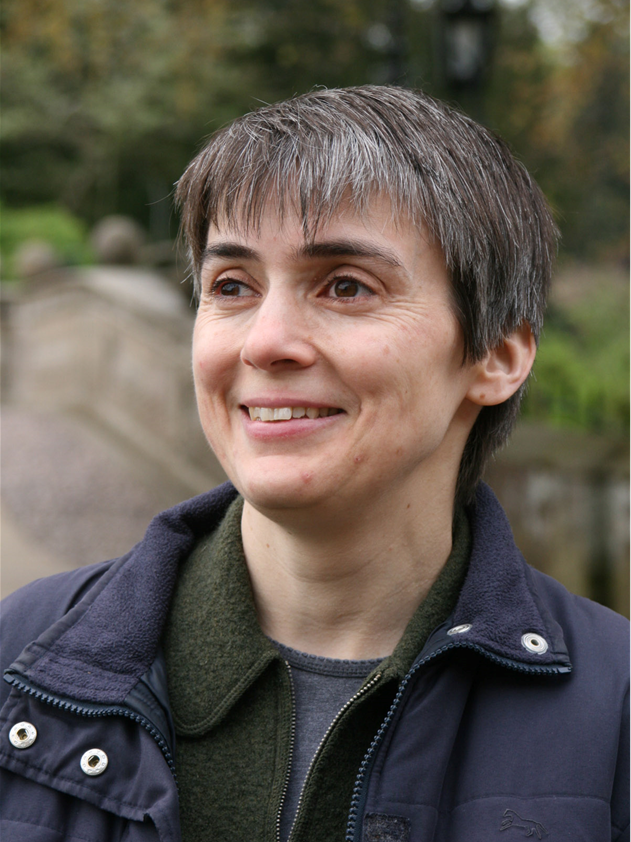
Ottoline Leyser, University of Cambridge, UK
Auxin and the self-organisation of plant form
Monday, October 29 - 09:40 AM - 10:20 AM - AUDITORIUM LUMIERE
Plants are exquisitely environmentally responsive, dynamically adjusting their growth and development according to the prevailing environmental conditions. We are using the regulation of shoot branching as a model system to investigate the mechanisms underlying this developmental plasticity. Shoot branching is regulated by an environmentally sensitive network of systemically moving hormone signals, providing a rich source of information that can be locally interpreted to determine branching behaviour. At the heart of this network is the auxin transport system. All active shoot apices export auxin into the polar auxin transport stream, which transports it rootward. Thus ultimately all shoot apices are in communication through their export of auxin into shared auxin transport paths to the root. Competition between apices for these transport paths can explain a range of phenomena in shoot branching, such as apical dominance. The shoot auxin transport system involves diverse transporters and mounting evidence suggests that different transporters are differentially sensitive to shoot branching regulatory signals, including auxin flux, cytokinin and strigolactone. We are exploring the mechanistic basis and functional significance of this diversity.
Andrew Oates
Andrew Oates, École polytechnique fédérale de Lausanne, Switzerland
Clocks and timers in development
Sunday, October 28 - 11:50 AM - 12:30 PM - AUDITORIUM LUMIERE
After positions at the Max Planck Institute for Molecular Cell Biology and Genetics in Dresden, Germany, University College London and the Francis Crick Institute in the UK, Andy Oates recently joined École Polytechnique Fédéral de Lausanne (EPFL) in Switzerland where he is the head of the Timing, Oscillation, Patterns Laboratory. The group studies a population of coupled genetic oscillators in the vertebrate embryo termed the segmentation clock. This system drives the rhythmic, sequential, and precise formation of embryonic body segments, exhibiting rich spatial and temporal phenomena spanning from molecular to tissue scales. Individual oscillating cells are synchronized with their neighbors, forming a coherent wave pattern of gene expression. How these wave patterns arise and how they are regulated during embryogenesis is not clear. I will describe recent progress in understanding the behavior of individual cells from the zebrafish as they slow their oscillations and differentiate during segmentation, and discuss how this gives rise to the tissue-level wave patterns. Central to this understanding is the concept of a timer that regulates the duration of a clock. This perspective reveals what part of the oscillatory cycle is changing as the cells slow and stop.
Barbara Prainsack
Barbara Prainsack, University of Vienna, Austria
Barbara Prainsack is a Professor at the Department of Political Science at the University of Vienna, and at the Department of Social Science, Health & Medicine at King’s College London. She is also a member of the European Group on Ethics in Science and Technologies advising the European Commission. Her current research focuses on personalised and “precision” medicine, on citizen participation in science and medicine, and the role of solidarity in medicine and healthcare. Her presentation will draw upon her latest books Personalized Medicine: Empowered Patients in the 21st Century? (NYU University Press, 2017)
Is precision medicine “open”?
Wednesday, October 31 - 05:50 PM - 06:30 PM - AUDITORIUM LUMIERE
The meaning of personalised medicine has shifted in recent years. Current iterations of personalisation as precision medicine make three key promises: First, to go beyond genomic information to include wider and more diverse datasets including non-molecular data; second, to overcome the “actionability gap” between insights obtained at the population level and the applicability of such knowledge at level of individual patients; and third, to move from symptomatic and episodic to continuous and presymptomatic medicine. In the context of such an understanding of precision medicine that hinges on large amounts of digital data, “openness” is typically understood as policies and practices that make “data sharing” possible and thus ensure the availability of the very datasets required for precision medicine in the first place. I argue that such an understanding of openness is unduly simplistic. Instead, openness in the context of personalized medicine should be understood in a threefold way: (a) in an ontological sense, pertaining to the openness of the category of the “person” in personalised medicine; (b) in a pluralistic sense, pertaining to the plurality of personal and societal values informing medical decision making; and (c) in an emancipatory sense, counteracting concentrations of power around new corporate actors – including consumer tech companies – in the health domain.
Marie-Anne Félix
Marie-Anne Félix, Institute of Biology of the Ecole Normale Supérieure, Paris, France
Marie-Anne Félix is a CNRS Research Director in the Institute of Biology at the Ecole Normale Supérieure in Paris. Her research encompasses quantitative and evolutionary biology of vulva development in C. elegans and other nematode species and studies of natural populations of these species, including their population genetic structure and natural pathogens.
Variational properties in a developmental system with six precursor cells
Tuesday, October 30 - 09:40 AM - 10:20 AM - AUDITORIUM LUMIERE
A key question in evolutionary biology is the extent to which the process of phenotype construction biases and constrains phenotypic evolution. In other words, what is the effect of random mutation at the phenotypic level? Do directions of phenotypic space with low robustness to random mutation correspond to directions of fast evolution?
The development of the vulva in the nematode Caenorhabditis elegans provides a model system where the fates and variational properties of each of six precursor cells can be compared quantitatively. We thus examined their properties of robustness to noise, environmental perturbations and spontaneous mutation, and their evolutionary variation. We find that:
- The central pattern of 2°-1°-2° vulval fates is highly robust to various environmental perturbations, but the developmental system leading to this invariant pattern evolves.
- The fate of the most anterior cell, P3.p, is the most sensitive to various perturbations (stochastic noise, environmental variations and spontaneous mutation accumulation), and is also that evolving most in the Caenorhabditis genus.
We further sought to understand the developmental basis for these differential variational properties of the six cells. Although most of the key signaling pathways (EGF, Wnt and Notch) involved in specification of the cell fates have now been identified, still very little is understood in quantitative terms. Through pathway dosage modulation and single molecule FISH hybridisation, we investigated quantitative aspects of system behaviour by experimentally perturbing the three pathways. The relative frequency of specific variants in the environmental perturbation and spontaneous mutation experiments above correspond to measurable properties of the developmental system, such as sensitivity to the dose of the relevant signaling molecules (EGF, Notch and Wnt pathways).
In order to test whether the high mutational variance of P3.p cell fate was due to a few loci with a particularly high mutation rate, we characterized the molecular nature of causal mutations. Only one of these genes is an obvious Wnt pathway gene, suggesting that mutations in a large number of genes may affect P3.p.
Overall, these results indicate that the low robustness of a trait to random mutation may produce evolutionary trends at the phenotypic level.
Mariko Okada
Mariko Okada, Osaka University, Japan
Mariko Okada (Hatakeyama) is a professor of Osaka University, Japan. She has been in systems biology field for about 18 years, with particular focus on signaling network in cancer and immune systems. For recent years, she is more interested in signaling outcomes, such as transcription, epigenetics and metabolism and tries to understand this whole system in a quantitative and data-driven manner.
Quantitative transcription control mediated by signaling network
Monday, October 29 - 05:50 PM - 06:30 PM - AUDITORIUM LUMIERE
Cellular fate decision is a time-development process and regulated by sequential activation of transcription factors. Such transcriptional regulation is closely associated with the epigenetic changes of chromatin structure, histone modifications and DNA methylation. Recently, super-enhancer (SE) has been suggested to determine cell specificity through generation of a threshold gene expression response, quantitatively different from the graded response controlled by typical-enhancers (TEs). To understand this mechanism, we try to identify a quantitative relationship among chromatin status, SE formation and SE-mediated transcription from integrative analysis of next-generation sequence (NGS) data by focusing on signaling-medicated NF-κB (RelA) gene regulation. Our analysis shows that chromatin opening is essential for SE formation, significant NF-κB binding and switch-like gene expression of NF-κB target genes, whereas the chromatin closing induced the loss of SE, NF-κB dissociation and steep gene down-regulation. A kinetic model could explain the allosteric Hill-type responses of SE and TE-medicated transcription by transcription factor binding. The study indicates that the degree in open-close chromatin status selects the enhancer modes and that is necessary for signal-dependent threshold/graded gene expression to determine the cell fate.
Takashi Hiiragi
Takashi Hiiragi, European Molecular Biology Laboratory, Heidelberg, Germany
Graduated from Kyoto University in 2000, Takashi Hiiragi started his group since 2002 at Max-Planck Institute of Immunobiology in Freiburg after his postdoc work with Davor Solter. He was an independent group leader at Max-Planck Institute for Molecular Biomedicine in Muenster, before moving to EMBL Heidelberg in 2011. A recipient of the ERC Starting and Advanced Grants. Looking at the molecular, cellular and systems levels, the Hiiragi group studies how, early in mammalian development, the embryo is self-organised from a spherical mass of cells.
Self-organisation in mouse development
Monday, October 29 - 09:00 AM - 09:40 AM - AUDITORIUM LUMIERE
A defining feature of living systems is the capacity to break symmetry and generate well-defined forms and patterns through self-organisation. Our group aims to understand the principle of multi-cellular self-organisation using early mouse embryos as a model system. Mammalian eggs lack polarity and symmetry is broken during early embryogenesis. This results in the formation of a blastocyst consisting of three cell types, each distinct in its position and gene expression. Our studies revealed that morphogenesis and gene expression are highly dynamic and stochastically variable during this process. Determining which signal breaks the symmetry and how the blastocyst establishes a reproducible shape and pattern despite the preceding variability remains fundamental open questions. We have recently developed an experimental framework that integrates biology, physics and mathematics. We aim to understand how molecular, cellular and physical signals are dynamically coupled across the scales for self-organisation during mammalian development.
Uwe Sauer
Uwe Sauer, Institute of Molecular Systems Biology, Zurich, Switzerland
As a trained microbiologist and with a postdoc in metabolic engineering, Uwe’s research as a PI and since 2006 as a professor for systems biology at the ETH Zurich focused on fundamental questions related to microbial metabolism. His lab combines modeling with quantitative experimentation, in particular mass spectrometry-based methods for 13C-flux analysis and high-throughput metabolomics. His main focus presently is on understanding how microbes coordinate their metabolic operation on short and long time scales.
Metabolic coordination through metabolite-protein interactions
Wednesday, October 31 - 09:40 AM - 10:20 AM - AUDITORIUM LUMIERE
How do bacteria know what goes on in their environment and how to they make appropriate decisions? While some bona fide extracellular sensors are known, there are far more environmental conditions and cellular responses than could possibly be dealt with through dedicated sensors. Instead, most microbial responses are based on intracellular changes to environmental changes. One of the first affected networks to just about any extracellular change is metabolism that passively responds to nutritional or chemical/physical challenges. Since fluxes and intracellular metabolite levels respond within seconds, allosteric binding of metabolites to regulatory proteins and enzymes is a highly effective and rapid sensing mechanism. Different from well-establish methods to assess physical interaction between proteins and between proteins and nucleic acids, however, methods to assess metabolite-protein interactions are still in their infancy. At present we know on the order of 1500 unique regulatory metabolite-protein interactions (1), which is only the tip of the iceberg (2). Beyond knowing the interaction topology, I will focus in this talk on the even more challenging and conceptual problem: understanding which of the many regulation mechanisms actually matter for a given adaptation to elicit an appropriate physiological system response. The surprising result for E. coli is that only very few regulation events appear to be required for a given transition, typically involving less than a handful of active regulators (3).
- Reznik, Christodoulou, Goldford, Briars, Sauer, Segre & Noor. Cell Reports 20: 2666-2677 (2017).
- Piazza, Kochanowski, Cappelletti, Fuhrer, Noor, Sauer & Picotti. Cell 72:358-372 (2018).
- Kochanowski, Gerosa, Brunner, Christodoulou & Sauer. Molecular Systems Biology 13: 903 (2017).
James Ferrell
James E. Ferrell, Jr., Stanford Center for Systems Biology, USA
Trigger waves in cellular regulation
Sunday, October 28 - 05:50 PM - 06:30 PM - AUDITORIUM LUMIERE
Trigger waves are self-regenerating, propagating fronts of activity that can spread without slowing down or losing amplitude. Familiar biological examples include action potentials and calcium waves; examples from everyday life include the spread of a fire through a forest, a virus through a population, or a meme through the internet. The minimal ingredients needed for a trigger wave are bistable chemical reactions plus some sort of spatial coupling mechanism.
Here I will present our recent studies that show that two very different phenomena—mitosis and apoptosis—are brought about by regulatory systems whose activation spreads through the cytoplasm via trigger waves. The proteins and biochemical processes involved are different, but the underlying logic is the same.
James Sharpe
James Sharpe, The European Molecular Biology Laboratory, Barcelona, Spain
Limb development, Turing patterns, and Computer modelling
Tuesday, October 30 - 09:00 AM - 09:40 AM - AUDITORIUM LUMIERE
James Sharpe was originally captivated by computer programming, but upon learning about the digital nature of the genetic code, chose to study Biology for his undergraduate degree at Oxford University (1988-1991). He then did his PhD on the genetic control of embryo development at NIMR, London (1992-1997) and in parallel started writing computer simulations of multicellular development. During his post-doc in Edinburgh, he began modelling the dynamics of limb development, and also invented a new optical imaging technology called Optical Projection Tomography (OPT), which is dedicated to imaging specimens too large for microscopy - tissues and organs. In 2006 he moved to Barcelona, becoming a senior group leader at the Centre for Genomic Regulation, and focusing on a systems biology approach to modelling limb development – combining experimentation with computer modelling. In this way the group demonstrated that the signalling proteins which pattern the fingers during embryogenesis, act as a Turing reaction-diffusion system. In 2011 he became the coordinator of the Systems Biology Program, and in 2017 was recruited to EMBL as Head of the new Barcelona outstation on Tissue Biology and Disease Modelling.
Ines Thiele
Ines Thiele, Luxembourg Centre for Systems Biomedicine and University of Luxembourg
Ines Thiele is the group leader of the Molecular Systems Physiology group at the Luxembourg Centre for Systems Biomedicine and an Associate Professor for Systems Biomedicine at the University of Luxembourg. Her research aims to improve the understanding of how diet influences human health. Therefore, she uses a computational modeling approach, termed constraint-based modeling, which has gained increasing importance in systems biology. Her group builds comprehensive models of human cells and human-associated microbes; then employs them together with experimental data to investigate how nutrition and genetic predisposition can affect one's health. In particular, she is interested in applying her computational modeling approach for better understanding inherited and neurodegenerative diseases.
Personalized whole-body models integrate metabolism, physiology, and the gut microbiome
Wednesday, October 31 - 09:00 AM - 09:40 AM - AUDITORIUM LUMIERE
Precision medicine relies on the availability of realistic, mechanistic models that capture the complexity of the human body. Comprehensive computational models of human metabolism have been assembled by the systems biology community, which summarize known metabolic processes occurring in at least one human cell or organ. However, these models have not yet been expanded to connect with whole-body level processes. To address this shortcoming, we built whole-body metabolic models of a male (deemed Harvey) and a female (deemed Harvetta) starting from the existing human metabolic models, physiological and anatomic information, comprehensive proteomic and metabolomic data, as well as biochemical data obtained from an extensive manual literature review. We tested the predictive capabilities of the resulting whole-body metabolic models against the current knowledge of organ-specific and inter-organ metabolism. The final models contain 28 organs. Importantly, these whole-body models can be expanded to include the strain-resolved metabolic models of gut microbes. By parameterizing the whole-body metabolic models with physiological and metabolomic data, we connected physiology with molecular-level processes through networks of genes, proteins, and biochemical reactions. As a sample application of the whole-body metabolic models, I will demonstrate how different microbial composition leads to differences in host metabolism, such as the capability to produce important neurotransmitters in the brain and flux through liver enzymes, with implications for the gut-brain axis as well as for microbiome-mediated liver toxicity. The predictions were consistent with our current understanding but also highlighted that different microbiota composition can lead to high inter-person variability. I envisage the microbiome-associated whole-body metabolic models will usher in a new era for research into causal host-microbiome relationships and greatly accelerate the development of targeted dietary and microbial intervention strategies.
Mustafa Khammash
Mustafa KHAMMASH, ETH Zurich, Switzerland
Mustafa Khammash is the Professor of Control Theory and Systems Biology at the Department of Biosystems Science and Engineering at ETH Zurich, Switzerland. He works in the areas of control theory, systems biology, and synthetic biology. His lab develops theoretical, computational, and experimental methods aimed at understanding the role of dynamics, feedback, and randomness in biology. He is currently developing new theoretical and experimental approaches for the design of biomolecular control systems and for their realization in living cells.
Prof. Khammash received his B.S. degree from Texas A&M University in 1986 and his Ph.D. from Rice University in 1990, both in electrical engineering. In 1990, he joined the engineering faculty of Iowa State University, where he created the Dynamics and Control Program and led the control group until 2002. He then joined the engineering faculty at the University of California, Santa Barbara (UCSB), where he was Director of the Center for Control, Dynamical Systems and Computation (CCDC) until 2011 when he joined ETH Zurich. He is a Fellow of the IEEE, IFAC, and the Japan Society for the Promotion of Science (JSPS).
Rationally-Designed Biomolecular Control Systems
Thursday, November 1 - 09:00 AM - 09:40 AM - AUDITORIUM LUMIERE
Nature relies on feedback control systems to achieve regulation at every level of biological organization. Such systems are also ubiquitous in man-made machines and devices. Can these systems be rationally designed and genetically engineered into living cells where they would act as autonomous molecular controllers that robustly steer the cell's behavior? This is the main topic of my presentation. I will start by explaining the main challenges facing the implementation of control systems in living cells. I then present a theoretical framework for the design and synthesis of molecular controllers that achieve robustness-by-design in the noisy environment of the cell. Guided by this theory, I will demonstrate a novel designer gene network that uses integral feedback to achieve robust perfect adaptation in bacterial cells. I will illustrate the tunability and robustness properties of this synthetic network and will highlight its potential use as a universal controller for regulation of biological variables in arbitrary networks. Finally, I will discuss the potential impact of biomolecular control systems in industrial biotechnology and medical therapy.
Nathalie Balaban
Nathalie Q. Balaban, The Hebrew University of Jerusalem, Israel
Nathalie Balaban completed her PhD in Condensed Matter Physics. Since her post-doctoral research, she became interested in cell-to-cell variability, focusing on the response of bacteria to antibiotics. Her work has explored the genetic networks that amplify noise to generate cell-to-cell variability, and how variability of growth dynamics can evolve to match the time-scale of environmental changes. The results shed new light on how treatment failure may evolve and lead to antibiotic resistance. She is a member of the European Academy of Microbiology and a Fellow of the American Academy of Microbiology.
To grow or not to grow: Phenotypic variability of growth in single cells
Thursday, November 1 - 09:40 AM - 10:20 AM - AUDITORIUM LUMIERE
Keynote speakers
Johan Elf

Johan Elf, Uppsala University, Sweden
Adventures with single molecules in bacterial cells
Thursday, November 1 - 10:50 AM - 11:50 AM - AUDITORIUM LUMIERE
I will present computational and experimental methods that we have developed to study dynamic intracellular processes at high spatial and temporal resolution in living cells. This includes methods for tracking individual molecules as well as methods for inferring intracellular binding states and reaction rates from these data. Both the experiments and analysis are optimised through simulated microscopy experiments based on rigorous stochastic modelling of the underlying reaction diffusion processes as well as the physics of the detection system.
With respect to biological systems, I will present our work on how DNA binding proteins find their specific targets in the chromosomal DNA and how the bacterial cell coordinates replication and cell division. Finally I will share results from our ongoing development of methods to bridge the gap between biophysics and genomics.
Julie A. Theriot
Julie A. Theriot, University of Washington, USA
Julie A. Theriot has recently moved to the University of Washington in Seattle, following 21 years at Stanford University. She is an investigator of the Howard Hughes Medical Institute. The experimental work of her research group focuses on quantitative measurement of the dynamic and mechanical behavior of structural components in living cells, exploring the molecular and biophysical mechanisms of various forms of cell motility and shape determination across a variety of eukaroytic and bacterial cell types. At ICSB 2018, her presentation will focus on large-scale mechanical integration in rapidly crawling cells.
The fast and the furious: Dissecting the mechanics and dynamics of rapid cell locomotion
Sunday, October 28 - 10:50 AM - 11:50 AM - AUDITORIUM LUMIERE
Directed crawling motility of animal cell types ranging from neurons to macrophages requires the coordinated force-generating activity of multiple mechanical elements. Much molecular detail is known about the constituents of some mechanical submachines such as the polymerizing actin network and the adhesion complexes, but it is not yet clear how these elements all work together to generate coherent, directed motion at the level of the whole cell. In order to understand cellular mechanisms of large-scale coordination, our work focuses on two extremely fast-moving cell types, the fish epidermal basal keratocyte responsible for the rapid closure of wounds in fish skin, and the human neutrophil that hunts down and kills microbial invaders. Under normal culture conditions, motile keratocytes assume an unusually simple, stereotypical, fan-like shape with a very large, thin lamellipodium densely packed with actin filaments, and can move persistently with nearly constant shape and speed. In contrast, neutrophils change shape dramatically during normal motion and exhibit significant intracellular fluid flows as well as complex cytoskeletal dynamics. We use a variety of quantitative methods based on live cell videomicroscopy to track the dynamic behavior of cytoskeletal elements and organelles as these cells move and respond to signals and obstacles in their environments including chemoattractants, electric fields, and physical variations such as changes in substrate adhesivity and microfabricated barriers. In parallel, we develop mathematical models that describe simplified physical frameworks for intracellular mechanics and dynamics in ways that can make quantitative predictions of experimental outcomes, which can then be used iteratively to refine the mathematical models. Despite their very different biological roles and behaviors, we find that keratocytes and neutrophils appear to share fundamental mechanisms of self-organization and movement coordination.