Keynote speakers
Johan Elf

Johan Elf, Uppsala University, Sweden
Adventures with single molecules in bacterial cells
Thursday, November 1 - 10:50 AM - 11:50 AM - AUDITORIUM LUMIERE
I will present computational and experimental methods that we have developed to study dynamic intracellular processes at high spatial and temporal resolution in living cells. This includes methods for tracking individual molecules as well as methods for inferring intracellular binding states and reaction rates from these data. Both the experiments and analysis are optimised through simulated microscopy experiments based on rigorous stochastic modelling of the underlying reaction diffusion processes as well as the physics of the detection system.
With respect to biological systems, I will present our work on how DNA binding proteins find their specific targets in the chromosomal DNA and how the bacterial cell coordinates replication and cell division. Finally I will share results from our ongoing development of methods to bridge the gap between biophysics and genomics.
Julie A. Theriot

Julie A. Theriot, University of Washington, USA
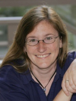
Julie A. Theriot has recently moved to the University of Washington in Seattle, following 21 years at Stanford University. She is an investigator of the Howard Hughes Medical Institute. The experimental work of her research group focuses on quantitative measurement of the dynamic and mechanical behavior of structural components in living cells, exploring the molecular and biophysical mechanisms of various forms of cell motility and shape determination across a variety of eukaroytic and bacterial cell types. At ICSB 2018, her presentation will focus on large-scale mechanical integration in rapidly crawling cells.
The fast and the furious: Dissecting the mechanics and dynamics of rapid cell locomotion
Sunday, October 28 - 10:50 AM - 11:50 AM - AUDITORIUM LUMIERE
Directed crawling motility of animal cell types ranging from neurons to macrophages requires the coordinated force-generating activity of multiple mechanical elements. Much molecular detail is known about the constituents of some mechanical submachines such as the polymerizing actin network and the adhesion complexes, but it is not yet clear how these elements all work together to generate coherent, directed motion at the level of the whole cell. In order to understand cellular mechanisms of large-scale coordination, our work focuses on two extremely fast-moving cell types, the fish epidermal basal keratocyte responsible for the rapid closure of wounds in fish skin, and the human neutrophil that hunts down and kills microbial invaders. Under normal culture conditions, motile keratocytes assume an unusually simple, stereotypical, fan-like shape with a very large, thin lamellipodium densely packed with actin filaments, and can move persistently with nearly constant shape and speed. In contrast, neutrophils change shape dramatically during normal motion and exhibit significant intracellular fluid flows as well as complex cytoskeletal dynamics. We use a variety of quantitative methods based on live cell videomicroscopy to track the dynamic behavior of cytoskeletal elements and organelles as these cells move and respond to signals and obstacles in their environments including chemoattractants, electric fields, and physical variations such as changes in substrate adhesivity and microfabricated barriers. In parallel, we develop mathematical models that describe simplified physical frameworks for intracellular mechanics and dynamics in ways that can make quantitative predictions of experimental outcomes, which can then be used iteratively to refine the mathematical models. Despite their very different biological roles and behaviors, we find that keratocytes and neutrophils appear to share fundamental mechanisms of self-organization and movement coordination.

