Partenaires
Rechercher
Accueil du site > Thèmes de recherche > Dynamics of swimming bacteria in the vicinity of surfaces. Project leader : Laurence Lemelle.
Dynamics of swimming bacteria in the vicinity of surfaces. Project leader : Laurence Lemelle.
- Lemelle L. (USR3010 & UMR5570 ), Chatre E. (USR 3010), Haftek Z. (USR3010) et Place C. (USR3010 & UMR5672)
- Joint Work With :Jean-Francois Palierne and Cédric Vaillant, Laboratory of Physics,UMR 5672 CNRS/ENS de Lyon.
Planktonic bacteria reach mineral or biological surfaces in their environments either through passive transport by convection-diffusion or gravity or, more efficiently, by active propulsion using flagella. On surfaces, the bacterial motility persists in a drastically modified form. Dark field videomicroscopy experiments were developed to statistically quantify this change. A Hydrodynamic model allowed to identify the critical parameters for the bacterial propulsion in the vicinity of surface. Tracking of bacteria in dark-field videomicroscopy at low magnification allows observation of a large number of bacteria under physiological conditions. The protocols for culture of E. coli and for optical imaging techniques in dark field contrast near a planar surface were established. A drop of bacterial cells solution deposited on a glass lamella is observed using an inverse microscope with an EBCCD Hamamatsu camera. This method allows observations of the cells with a strong and constant contrast during the experiment without modifying the dynamics of the cellular swimming (Fig. 1). The trajectories are determined using a data treatment developed at the Joliot-Curie lab, recording the bacterial geometry and their dynamics properties (local velocity, average or median velocity, and radius of curvature).
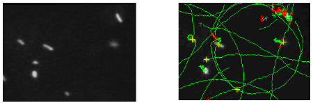
Figure 1a : Dark field image of E. coli cell on a glass lamella surface recorded with a x40 objective. Figure 1b : Image of the calculated trajectories (en vert) for the bacteria observed on the previous figure.
Near a solid surface, E. coli becomes trapped and swims in circles. Tracking of bacteria in dark-field videomicroscopy evidences a direct relation between curvature radius of the trajectory and bacterium speed. A hydrodynamic model for this motion on a non-slipping surface was proposed by E. Lauga & al. [Biophys. J. 2006] using the resistive force approximation with moderate agreement with observations. A fair agreement obtains with a revised version of Lauga’s model. Models predict the nanometric distance of the cell to the surface is the critical hydrodynamic parameter. A TIRF LEICA microscope, acquired thanks to a "mi-lourd" support from CNRS, was installed in the Laboratoire Joliot Curie at the end of 2007. Different strategies of physiological fluorescent staining were explored. Finally, bacterial mutants bearing a plasmid that encode for a periplasmic protein fused to mCherry (a red fluorescent protein) were produced and demonstrated the same swimming characteristic as the wild type. Preliminary TIRF videomicroscopy experiments (Figure 2a) with bacteria revealed a nanometric range for the distance of the body to the surface (10 nm < d < 300 nm).
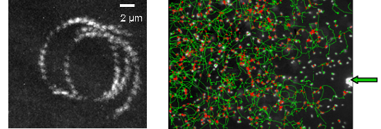
Figure 2a : trajectory of E. coli swimming close to a surface observed by TIRF microscopy (magnification X60, staining Syto). Figure 2b : Trajectories of E. coli (dark field magnification X20) swimming close to a surface while a chemorepellent solution is microinjected on the surface (extremity of the capillary is indicated by a green arrow).
Since hydrodynamics predicts a physical trapping near surfaces, E. coli cells are predicted to be loosely capable to orientate their swimming versus chemicals. Chemo-repellent solutions were microinjected within few m of the surface (Figure 2b). Observations are all consistent with a negative chemotaxis response of cells moving down a spherical repulsive-gradient. The cellular distribution has a radial symmetry centered on the aperture of the capillary. The number of motile cells and their median speed decrease and reach minimal values in less than 100s. Once the capillary is removed, the system recovers its initial state. Responses of the tsr(-) and tar(-) mutants to Ni-microinjection revealed the implication of the MCP receptors. Herein swimming cells can sense surface chemical environments and overcome hydrodynamics laws to move toward preferential environments. Other techniques to induce chemical gradients must be now explored to reach a satisfactory reproducibility.
This work was supported by iterative private subcontracting since 2007 guaranteeing the expenses ( > 20 kEuros) and the salary of an engineer in microbiology.
Two publications are under submition :
- Radius of curvature of E. coli trajectories near a planar surface increases with the swimming speed. by Lemelle L., Palierne J-F., Chatre E. and C. Place.
- Resistive force theory with slip at the boundaries applied to bacterial locomotion in viscous media by Palierne J-F., Lemelle L., Chatre E. and C. Place


