
 Directory
Single Molecule Methods
The potential and need of single-molecules genomic methods:
To investigate complex biomolecular processes like replication, transcription, and DNA repair, we frequently rely on assays such as ChIP-seq for protein-DNA interactions, Hi-C or Micro-C for 3D genome organization, and MNase-seq or ATAC-seq for nucleosome structure. These methods provide valuable insights but have inherent limitations, particularly when analyzing multiple parameters in conjunction.
For example, analyzing ChIP-seq experiments of two proteins that bind the same locus often suggests coincident binding. However, because these findings arise from separate experiments, they cannot confirm simultaneous binding on the same DNA molecule; it’s possible that the proteins bind independently, competitively, or synergistically. Fully understanding such nuanced biological interactions requires techniques capable of detecting multiple proteins on the same unique molecule of DNA (single-molecule), something population-based methods are fundamentally unable to achieve.
Similarly, investigating relationships between protein binding, 3D genome architecture, and nucleosome positioning demands single-molecules techniques. Current approaches rely on aggregated, population-average data, which obscures molecular details essential for understanding these complex interactions. As the field progresses, new single-molecule and single-cell techniques will emerge to overcome these limitations, promising breakthroughs over the next decade.
One concrete illustration is microscopy experiments, which inherently represent single-cell approaches. Recent live-cell imaging has revealed that transcriptional activity at a gene locus decreases just before its replication (Tsirkas et al., 2022), a discovery made possible by simultaneously tracking replication and transcription. Previous techniques, such as BrdU-IP and RNA-seq, only provided population-level data, which masked these co-occurring events. Although microscopy overcomes this limitation, it also has drawbacks: only a limited number of cells (typically hundreds) and a single locus can be visualized at once. To map dynamic processes genome-wide, methods capable of capturing replication and transcription on millions of single molecules in one experiment are needed.
In the field of 3D genome interactions this led to the development of methods like SPRITE (Quinodoz et al., 2018) and ChIA-Drop (Zheng et al., 2019) that track chromatin interactions at the single-molecule level, but these methods remain costly, complex, and lack sufficient resolution. My postdoctoral work directly addresses this technological gap, resulting in the development of Proximity Copy Paste (PCP) (Delamarre et al., 2025).
The PCP method:
This project is based on the PCP method that can detect protein footprints on DNA (nucleosomes, transcription factors and tightly bound proteins) at base-pair resolution, the spacing of neighboring nucleosomes and chromatin interactions, all on the same molecule of DNA. This assay is very versatile and will be used and further optimized to detect other properties of chromatin at the single molecule level.
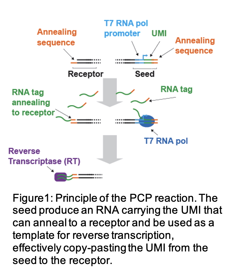
The principle of PCP relies on two molecular elements (fig.1): A Seed, which is a piece of DNA with a T7 RNA promoter, a UMI (Unique Molecular Identifier) and an annealing sequence. In the PCP reaction a T7 RNA polymerase will transcribes the seed and produce multiple copies of RNA carrying the UMI and annealing sequence information that will diffuse around the seeds. The second element is a Receptor molecule, comprising a 5’ end overhand harboring the annealing sequence. Receptor molecules in proximity to the seed will anneal to the RNA molecule that shares the complementary annealing sequence. The PCP reaction also utilizes a Reverse transcriptase that will use the RNA as a template and elongate the receptor molecule. As a result, the UMI that was initially present on the seed is now also present at the end of the receptor molecule. Importantly, a seed is not limited to one target and can tag several receptor molecules in the same reaction. The UMI is therefore copied from the seed and paste onto the receptor, hence copy/paste.
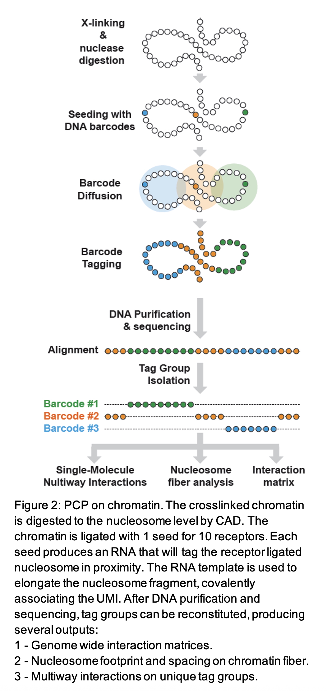
To conduct the PCP on chromatin (fig.2), the chromatin is fixed with formaldehyde and DSG before being digested by Caspase Activated DNase (CAD). Like the Micrococcal Nuclease (MNase), CAD does not digest protein protected DNA, mainly producing nucleosome size fragments. Once digested and end-repaired, the chromatin is ligated with a mixture of 1 to 10 seed to receptor ratio. While the ligation of seeds is not targeted, this will result in a distribution of the seeds on the chromatin, that can be determined after DNA sequencing. The PCP then takes place, and the seeds tag the receptor molecule in their proximity. Upon DNA purification and sequencing, tag groups can be reconstituted based on the UMI sequences. Nucleosomes that were in spatial proximity are tagged with the same UMI and footprinting, spacing, and long-range interaction can be identified.
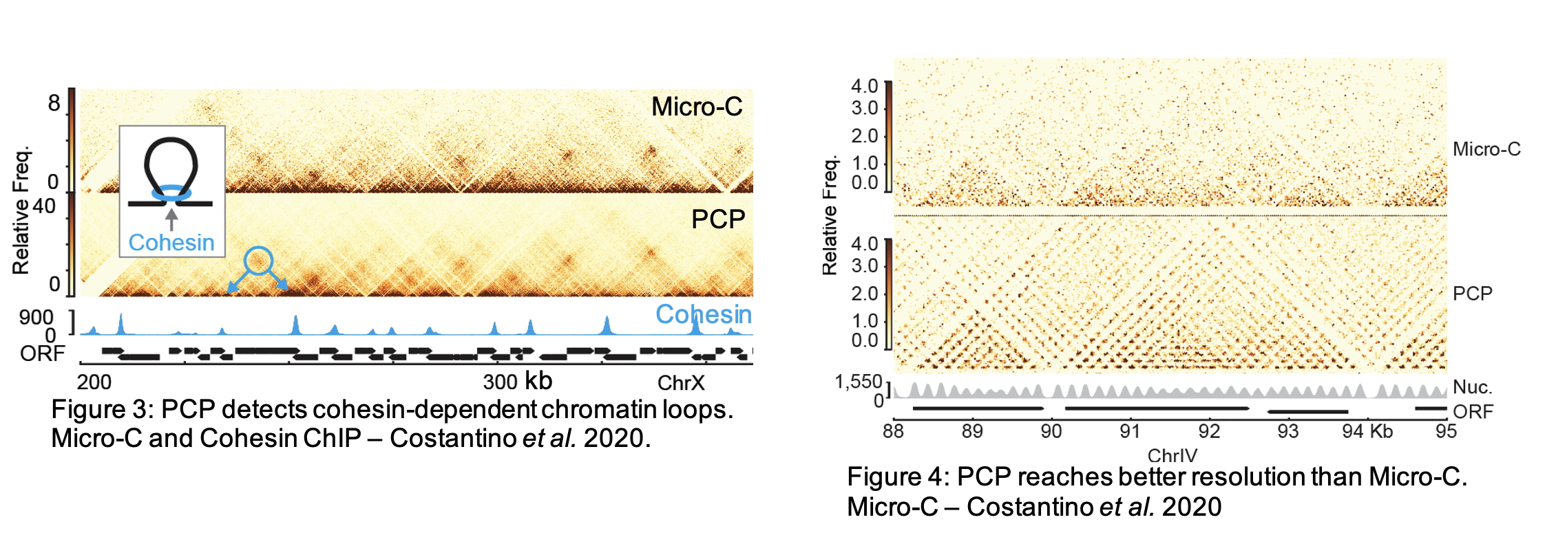
The classical way to represent DNA-DNA contacts is through interaction matrices (figs.3-4). In a matrix, each pixel represents a potential interaction between two points in the genome, while the intensity of the pixel represents the frequency at which each interaction was found. In figure 3 the blue circle highlights the interaction between two cohesin peaks. This represents the formation of chromatin loops by the ring shaped complex Cohesin (Costantino et al., 2020). PCP detects nucleosome connectivity at unprecedented resolution (fig4). While perfectible, PCP offers a better resolution, base pair foot-printing, detection of non-canonical nucleosome signals, detection of multiway interactions, and detection of proximal proteins out of DNA contact reach compared to Micro-C and Hi-C. In addition, by sequencing every nucleosome and not only the ones involved in contacts, PCP is also more quantitative than Micro-C or Hi-C.
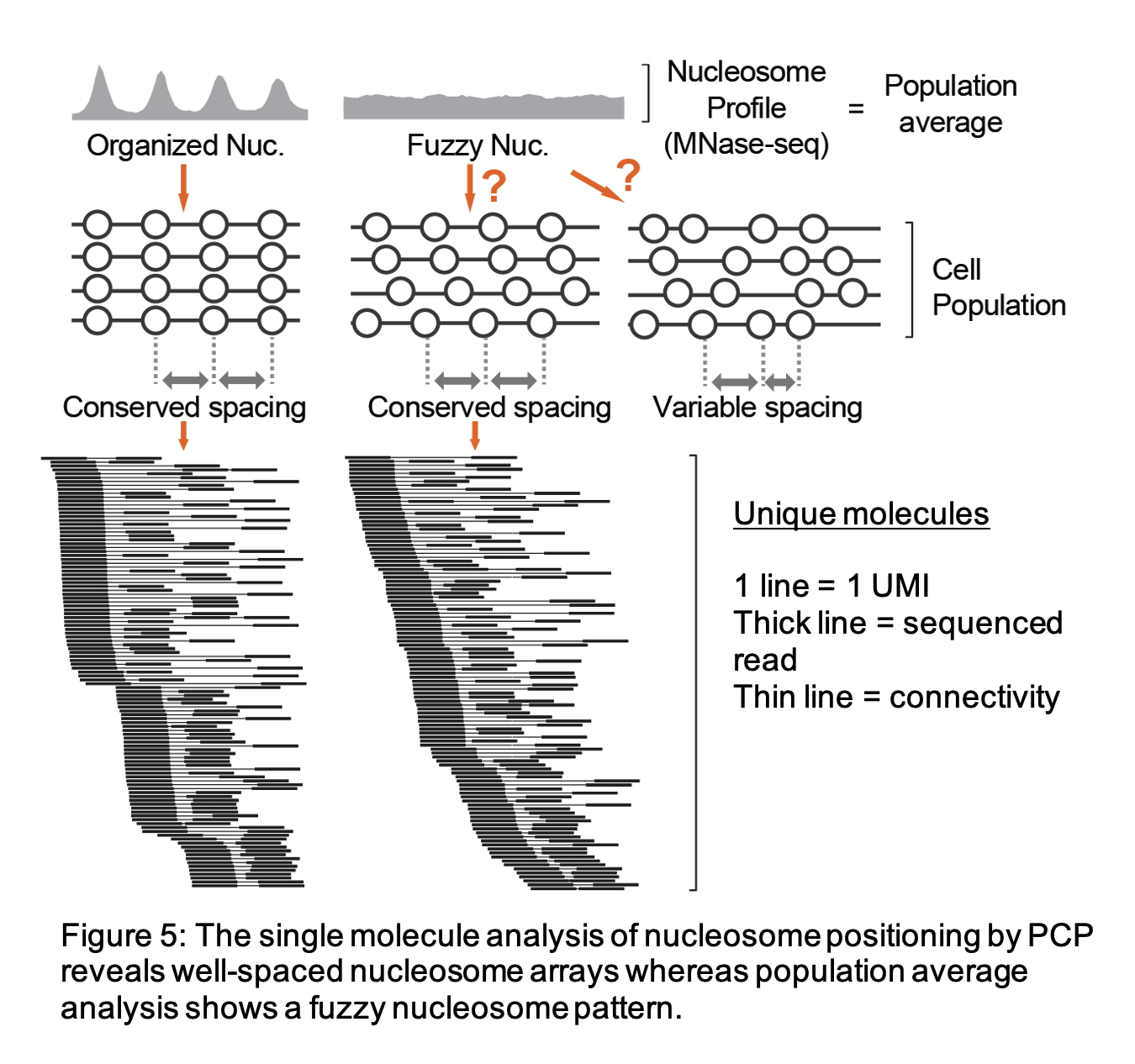
Importantly, PCP provides single-molecule resolution, allowing it to collect multiple parameters from the same unique DNA molecule. Chromatin fragments originating from the same DNA molecule are labeled with a unique UMI, enabling precise mapping of the positions of multiple nucleosomes and interactions on an individual DNA molecule. Single-molecule data can be represented as a line graph, with each line corresponding to a unique UMI; the thick lines represent the reads found and the thin line connecting them represent that these reads were linked.
The single-molecule analysis, as opposed to averaged readouts, offers distinct advantages. For instance, MNase-seq often yields a fuzzy nucleosome signal that could be interpreted in two ways — either as a highly disorganized nucleosome landscape or as a well-spaced nucleosome array with slight positional offsets between cells — single-molecule analysis, however, reveals a regularly spaced nucleosome array (fig5). This type of analysis also enables the identification of subpopulations within mixed signal populations, adding depth and clarity to the insights gathered.
References:
Costantino, L., Hsieh, T. S., Lamothe, R., Darzacq, X., & Koshland, D. (2020). Cohesin residency determines chromatin loop patterns. Elife, 9. https://doi.org/10.7554/eLife.59889
Delamarre, A., Bailey, B., Yavid, J., Koche, R., Mohibullah, N., & Whitehouse, I. (2025). Chromatin architecture mapping by multiplex proximity tagging. Mol Cell, 85(14), 2796-2811 e2795. https://doi.org/10.1016/j.molcel.2025.06.018
Quinodoz, S. A., Ollikainen, N., Tabak, B., Palla, A., Schmidt, J. M., Detmar, E., Lai, M. M., Shishkin, A. A., Bhat, P., Takei, Y., Trinh, V., Aznauryan, E., Russell, P., Cheng, C., Jovanovic, M., Chow, A., Cai, L., McDonel, P., Garber, M., & Guttman, M. (2018). Higher-Order Inter-chromosomal Hubs Shape 3D Genome Organization in the Nucleus. Cell, 174(3), 744-757 e724. https://doi.org/10.1016/j.cell.2018.05.024
Tsirkas, I., Dovrat, D., Thangaraj, M., Brouwer, I., Cohen, A., Paleiov, Z., Meijler, M. M., Lenstra, T., & Aharoni, A. (2022). Transcription-replication coordination revealed in single live cells. Nucleic Acids Res, 50(4), 2143-2156. https://doi.org/10.1093/nar/gkac069
Zheng, M., Tian, S. Z., Capurso, D., Kim, M., Maurya, R., Lee, B., Piecuch, E., Gong, L., Zhu, J. J., Li, Z., Wong, C. H., Ngan, C. Y., Wang, P., Ruan, X., Wei, C. L., & Ruan, Y. (2019). Multiplex chromatin interactions with single-molecule precision. Nature, 566(7745), 558-562. https://doi.org/10.1038/s41586-019-0949-1