


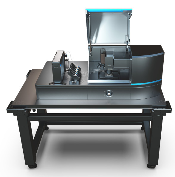
ENS de Lyon is the first institution in France to invest in the only instrument in the world that allows the simultaneous manipulation and visualization of interactions between single biomolecules in real time.
8 teams in 3 laboratories – LPENSL, LBMC and RDP – are now working with this instrument.
ENS de Lyon is the first institution in France to invest in the only instrument in the world – the C-Trap – which simultaneously and in real time allows for the manipulation and visualization of interactions between unique biomolecules. It combines high-resolution optical clamps, confocal fluorescence microscopy and advanced microfluidics, all in a truly integrated system.
A partnership has been set up between the company that markets this instrument and the School. The C-Trap will therefore be available 8 weeks a year so that the company can demonstrate it to other French laboratories.
What is an optical clamp?
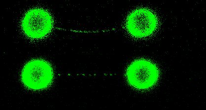
The optical clamp, or optical trap, is a tool that enables the trapping and manipulation of transparent microbeads but also biomolecules, cells, micro drops or organelles. It makes it possible to exert extremely weak forces, in the region of one piconewton (pN), comparable to the forces exerted naturally within our cells.
For this it uses the force resulting from the refraction of light, a laser beam. Multiple optical clamps, such as those acquired by the School, can even be used to simultaneously manipulate multiple objects and track their interactions.
Thanks to this technology, it is possible to study, for example, the viscoelastic properties of proteins, the flexibility and resistance to the twisting of DNA strands, or protein/DNA interactions. But many other applications exist, mainly in biology but also in chemistry and physics. At ENS de Lyon, 8 teams in 3 laboratories – LPENSL, LBMC and RDP – are now working with this instrument.
Why combine optical tweezers with microfluidics and confocal microscopy?
If the optical clamps are used to catch and manipulate the molecules of interest; laminar flow microfluidics make it possible to introduce, assemble and position reactants; Finally, confocal fluorescence imaging makes it possible to record biochemical reactions and movements of molecules in real time.