


Publication of the IGFL in the journal PNAS on March 19, 2024. News by CNRS Biology on April 12, 2024.
During embryonic development, motor neurons, located in the spinal cord, emit extensions - the axons - which must find their way to their targets - the muscle cells. These motoneurons, last link between electrical and mechanical signals in the locomotor system, trigger movement via their axons.
Using the model zebrafish, research carried out by a team at the Institute of Functional Genomics of Lyon (IGFL, CNRS/ENS de Lyon), has described two proteins which, like red and green beacons at the entrance to a harbor, guide the motor axon's path.
The biologists bioprinted a path made of these proteins to faithfully mimic the one observed in vivo. They demonstrated that motor axon placed in this in vitro pattern followed exactly the path traced by these "beacon" proteins.
This discovery opens up promising prospects for the regeneration of damaged nerves. The findings are published in the journal PNAS.
Significance
En route to their muscle targets, growing motor axons are guided by a combination of diffusible and contact-mediated cues that attract or repel axons. Notably, motor axon navigation is locally influenced by extracellular matrix signposts that pave their trajectories. The repertoire and function of these contact-mediated cues and their cellular source are still under investigation. Using zebrafish, we show that differentiating muscle progenitors represent an important source of extracellular matrix cues. Our study reveals that the attractive Collagen XV-B is organized in a permissive route delimited by the repulsive Tenascin C to allow accurate navigation of motor axons. Our findings provide insights into how extracellular matrix participates in axonogenesis with interest in nerve regenerative medicine for peripheral nerve injury.
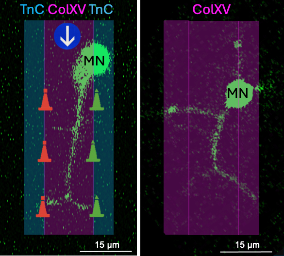
Credits: Laurie Nemoz-Billet and Florence Ruggiero
Illustration credits: Sandrine Bretaud and Florence Ruggiero
Reference
Dual topologies of myotomal collagen XV and Tenascin C act in concert to guide and shape developing motor axons. Laurie Nemoz-Billet, Martial Balland, Laurent Gilquin, Benjamin Gillet, Isabelle Stévant, Emilie Guillon, Sandrine Hughes, Gilles Carpentier, Elisabeth Vaganay, Frédéric Sohm, Vladimir Misiak, Mary-Julieth Gonzalez-Melo, Manuel Koch, Yad Ghavi-Helm, Sandrine Bretaud, and Florence Ruggiero. PNAS, March 19, 2024.
DOI : 10.1073/pnas.2314588121